Figure 2.
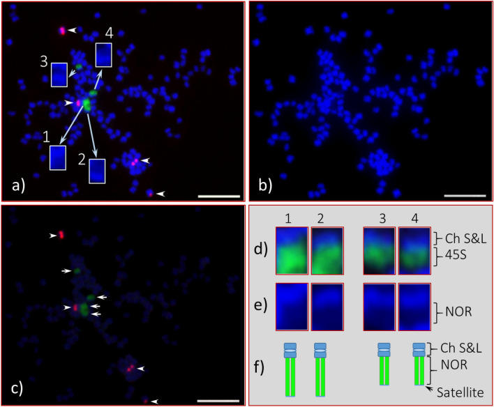
Fluorescent in situ hybridization (FISH) of 45S and 5S rDNA in somatic chromosome spreads of Adansonia digitata L. (Seedling-1). (a) Four 45S rDNA FISH signals (green) observed at the end of four chromosomes (also shown in “d”, enlarged image of each), and they are marked as 1, 2, 3, and 4 (inserts are enlarged DAPI stained chromosomes). (b) DAPI-stained chromosomes of the same cell as in “a”. (c) Same cell captured with reduced DAPI exposure that shows bright 45S (green, arrows) and 5S (red, arrowheads) rDNA signals. (d) Enlarged image of four 45S rDNA-bearing chromosomes with corresponding DAPI-stained chromosomes in “e”, depending on FISH signal intensities 1 and 2 are most likely a homologous pair and same for 3 and 4; (f) diagrammatic representation of these two homologous pairs of 45S rDNA bearing chromosomes. Scale bar is 5 µm.

