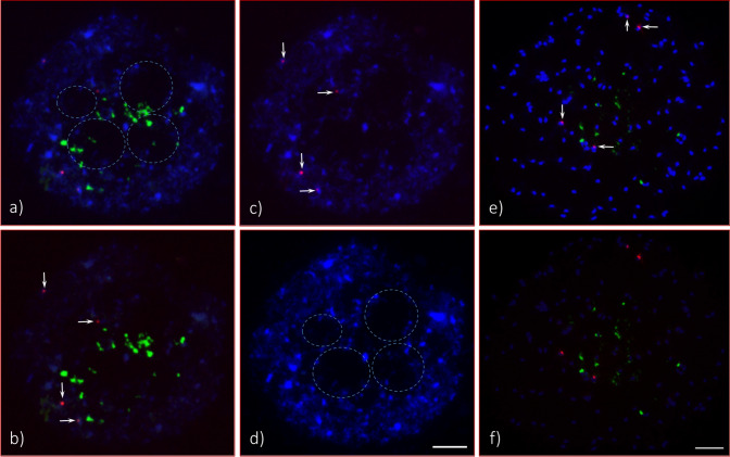
Figure 5.
Numerous 45S (green signals) and four 5S (red signals) observed in interphase and prophase cells of Adansonia digitata L. (Seedling-2). (a) As many as 30 + 45S rDNA FISH signals can be observed. (b) Same cell as in “a”, but the image was captured with short exposure time under DAPI filter to show bright green (45S rDNA) and red signals (5S rDNA, arrows). (c) Same cell as in “a”, showing 5S rDNA signals (arrows) in DAPI background interphase cell. (d) Same cell as in “a”, showing four hallow encircled areas, which are presumptive spaces for four nucleoli. (e) Late prophase cell with scattered green signals (45S rDNA) and four 5S rDNA signals (red, arrows) in DAPI background chromosomes. (f) Same cell as in “d”, about fifteen green signals (45S rDNA) and four red signals (5S rDNA) in reduced DAPI to show the brightness of the signals. Scale bars are 5 µm.

