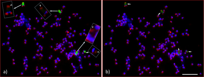
Figure 6.
Root tip chromosome spreads of Adansonia digitata L. (Seedling-1), analyzed with 45S rDNA and ATRS-type telomere oligonucleotide probes. (a) Four 45S rDNA (green) and telomere repeat (red) signals in DAPI-stained chromosomes background, inserts are the enlarged images of 45S rDNA-bearing chromosomes (with no green signals) showing the telomere signals at both ends of each chromosome, arrow heads show the telomere signals at the terminal end of each 45S rDNA signals. (b) Same cell as in “a” captured with short exposure time under FITC filter, the telomere signals at the terminal end of each 45S rDNA signals (light green signals) are brightly visible compared to “a”. Scale bar is 5 µm.

