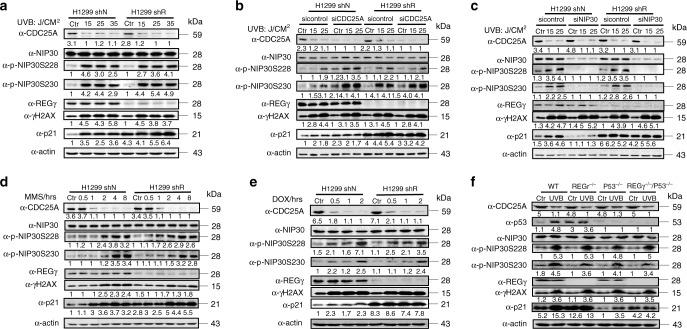
Fig. 5. The CDC25A–NIP30-REGγ pathway controls p21 following DNA damage.
a H1299 REGγ shN and shR cells were irradiated with 0, 15, 25, or 35 J/m2 UVB and harvested 12 h later. Cell lysates were analyzed by western blots with indicated antibodies. b H1299 REGγ shN and shR cells were treated with siRNA against CDC25A or a control siRNA followed by UVB irradiation. Cell lysates were subjected to western blot with indicated antibodies. c H1299 REGγ shN and shR cells were treated with siRNA against NIP30 or a control siRNA followed by UVB irradiation. Cell lysates were subjected to western blot with indicated antibodies. d, e CDC25A–NIP30 action after DNA damage by chemical reagents. H1299 REGγ shN and shR cells were treated with 0.2 mM MMS (d) or 10 μg/ml Doxorubicin (e) for the indicated time course. The levels of CDC25A, total NIP30, pNIP30Ser228, pNIP30Ser230, p21, REGγ, and γH2AX were determined by western blot. f In vivo action of the CDC25A–NIP30-REGγ pathway. REGγ+/+, REGγ−/−, P53−/−, and REGγ−/−/P53−/− newborn mice were exposed to UV light as described in “Methods”. Skin samples from exposed dorsal side or ventral side (un-exposed control) were examined for proteins indicated. All the experiments were repeated three times. Source data are provided in a Source Data file.

