Figure 1.

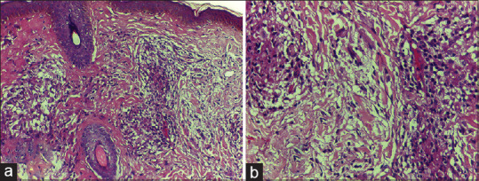
(a) Epithelioid granuloma showing intra-granuloma edema and dermal edema with separation of dermal collagen (H and E, ×200); 1 (b): high power view of Figure 1 (a)(H and E, x400)

(a) Epithelioid granuloma showing intra-granuloma edema and dermal edema with separation of dermal collagen (H and E, ×200); 1 (b): high power view of Figure 1 (a)(H and E, x400)