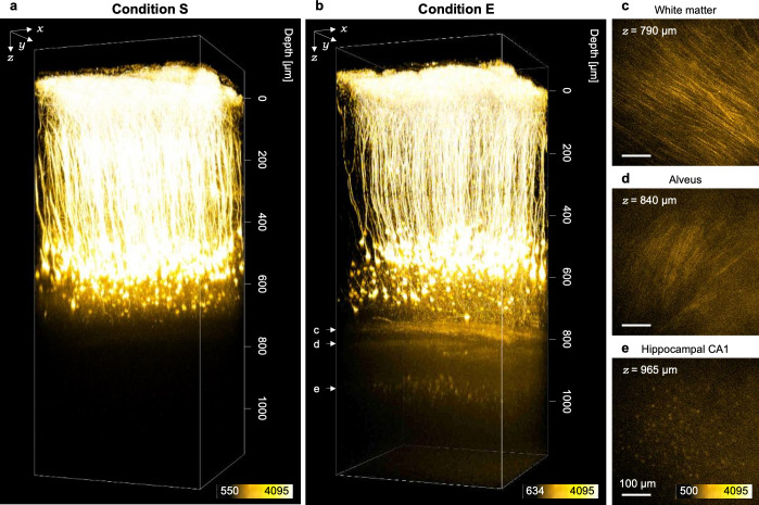
Fig 5. Two-photon imaging of cortical and hippocampal neurons under condition E.
(a, b) 3D reconstructed images of a living mouse brain obtained under (a) condition S and (b) condition E. Hippocampal CA1 neurons were clearly visualized in (b). (c–e) Fluorescent images of the white matter, the alveus of hippocampi and hippocampal CA1 neurons, respectively.

