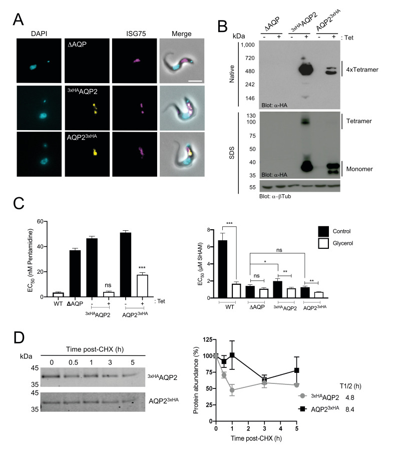
Fig 2. Characterisation of tagged TbAQP2.
A) Fluorescence microscopy of T. brucei 2T1 cells expressing tetracycline-regulated N- or C-terminal tagged AQP2 (3xHAAQP2 or AQP23xHA, respectively, in yellow). These proteins localise similar to ISG75 (magenta) at the flagellar pocket/endosomes. The triple aqp-null T. brucei 2T1 cells (ΔAQP) were also included as control. Scale bar 5 μm. B) Tet-regulated expression of N- or C-terminal HA-tagged AQP2. Both native-PAGE (upper panel) and SDS-PAGE (lower panel) αHA blots are shown. α–β tubulin was used as loading control. The presence of the different oligomeric species is indicated in the right-hand side of the panel. Note the presence of a high molecular weight form under SDS-PAGE in 3xHAAQP2 but not AQP23xHA. The triple aqp-null T. brucei 2T1 cells (ΔAQP) were also included as control. C) EC50 values for pentamidine (left panel) or salicylhydroxamic acid (SHAM; right panel) with or without 5 mM glycerol following expression of either 3xHAAQP2 or AQP23xHA. For multiparametric ANOVA, we compared the average values (n = 4 independent replicates) from wild type T. brucei 2T1 cells as reference for pentamidine, or from aqp-null cells for SHAM. * p<0.01, ** p<0.001, *** p<0.0001 from four independent replicates. D) Left panel; Representative western blotting (n = 3 independent replicates) from protein turnover assay monitored by cycloheximide (CHX) treatment in T. brucei 2T1 cells expressing either 3xHAAQP2 (upper panel) or AQP23xHA (lower panel). Right panel; Protein quantification from western blotting analysis in left panel for either 3xHAAQP2 (black square) or AQP23xHA (grey circles). Results are the mean ± standard deviation of three independent experiments (n = 3 independent replicates). The estimated half-life (t1/2) was calculated based on regression analysis using PRISM.

