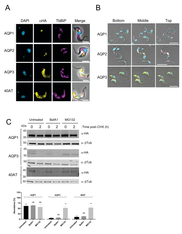
Fig 7. Differential turnover rate of the repertoire of AQPs in the bloodstream form of T. brucei.
A) Cell lines expressing N-terminal HA-tagged TbAQP1, TbAQP2, TbAQP3, field-isolate chimeric AQP2/3 (40AT) (Alexa Fluor 488; yellow) co-stained with the endoplasmic reticulum marker anti-BiP (magenta). DAPI (cyan) was used to label the nucleus and the kinetoplast. Scale bars, 5 μm. B) Three different confocal planes are shown for 2T1 cells expression TbAQP1, TbAQP2, or TbAQP3. The planes are defined from “Bottom” (far from the flagellar pocket) to “Top” (close to the flagellar pocket). Note a change in DAPI intensity as the images progress through the different planes. TbAQPs are denoted in yellow, DAPI in cyan, and TbISG75 in magenta. Scale bar, 10 μm. C) Upper panel; Representative western blot (n = 3 independent replicates) of protein turnover monitored by cycloheximide (CHX) treatment followed by pulse-chase assay. Cells were either untreated or treated with 100 nM of Bafilomycin A1 (BafA1) or 25 μM of MG132 for 1 h prior to harvest. Cells were harvested at 0 hours and 2 h post-CHX treatment and analysed by immunoblotting. Lower panel; Protein quantification representing the mean ± standard deviation of three independent experiments (n = 3 independent replicates). Statistical analysis was conducted using the signal from untreated cells at 2 h post-CHX treatment as reference group. * p<0.01, ** p<0.001, ns = not significant, using a t-test.

