
Figure 2. Brain MRIs finding obtained from 3 patients with C-NORSE.
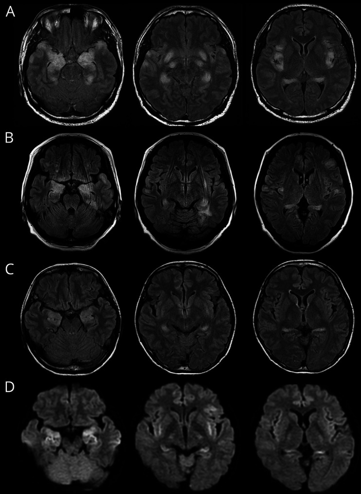
Brain MRIs show symmetric increased DWI or FLAIR signals in the amygdala, hippocampus, fimbria, claustrum, insula, and frontotemporal cortex. Basal ganglia and perisylvian opercular cortex are also involved in patients with C-NORSE (not shown). (A) A 37-year-old man; (B) a 49-year-old woman; (C and D) a 39-year-old woman; (A–C) FLAIR, (D) DWI. C-NORSE = cryptogenic new-onset refractory status epilepticus; DWI = diffusion-weighted image; FLAIR = fluid-attenuated inversion recovery.