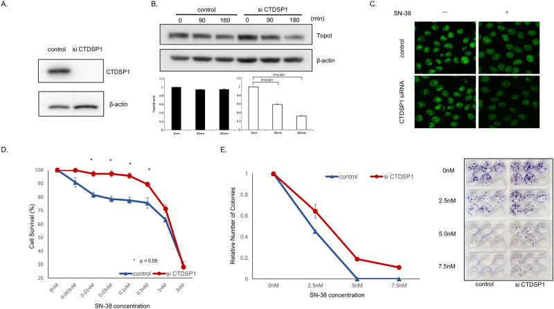
Fig 2. Silencing of CTDSP1 enhances topoI degradation and irinotecan resistance.
A, Cells transfected with CTDSP1 or control siRNA were lyzed and the cells’ lysates were immunoblotted with anti-CTDSP1 and anti-β-actin antibodies. B, HCT116-siRNA CTDSP1 or control siRNA, treated with 2.5 μM SN-38 were harvested after 90 and 180 min. Cell lysates were immunoblotted with anti-topoI and anti-β-actin antibodies. Cells’ lysates were immunoblotted with anti-topoI and anti-β-actin antibodies. C, EGFP was integrated with topoI in HCT116 cells using CRISPR/Cas9 system and CTDSP1 was knocked down in this cell line by siRNA. Cells were treated with 2.5uM SN-38 for 60 min and the topoI-EGFP signal was imaged by Leica SP5 confocal microscope. D, HCT116-siRNA-CTDSP1 or control siRNA were plated in a 96-well plate and treated with various concentrations of SN-38 for 72 h. Cell viability was determined by detecting the luminescence. E, HCT116-siRNA-CTDSP1 or control siRNA cells were plated in a 6-well plate and treated with various concentrations of SN-38 for 24 h. Then, 50 cells per well were plated in a 6-well plate. After 14 days, when colonies were apparent (right panel), colonies were counted and the relative number of colonies was determined (left panel).

