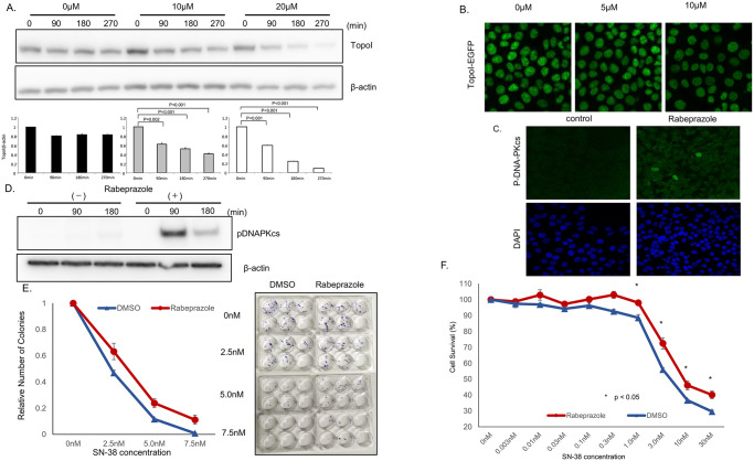
Fig 5. Rabeprazole promotes topo I degradation and irinotecan resistance.
A, HCT116 cells were plated in a 6-well plate and treated with various concentrations of rabeprazole (0, 10, 20 μM) for 72 h, and then with 2.5 μM SN-38 and harvested after 90 or 180 min. Cell lysates were immunoblotted with anti-topoI and anti-β-actin. B, Genomically edited HCT116 cells with TopoI-EGFP fusion proteins were treated with 5 and 10 μM of Rabeprazole for 48 hours and topoI-GFP protein level was analyzed by confocal microscope. C. HCT116 cells were plated in a 6-well plate, treated with 40 μM rabeprazole or DMSO for 72 h, and then with 2.5 μM SN-38, and harvested after 90 or 180 min. Cell lysates were immunoblotted with anti-pDNA-PKcs and anti-β-actin. D, HCT 116 cells were treated with rabeprazole, control and treated cells were analyzed by immunofluorescence analysis with anti-phospho-DNA-PKcs-pS2056 and confocal microscopy. E. HCT116 cells were plated in a 6-well plate and treated with rabeprazole or DMSO for 72 h. Then, 50 cells were plated in each well of a 6-well plate and treated with various concentrations of SN-38 for 24 hours. Cell colonies were counted after 14 days. F. HCT116 cells were plated in a 6-well plate and treated with 40 μM rabeprazole or DMSO for 72 h. Then, cells were plated in a 96-well plate and treated with various concentrations of SN-38 for 72 h. Cell viability was determined by luminescence detection.

