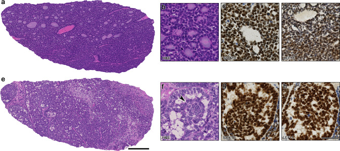
Fig. 1.
Morphological and immunohistochemical overview of the HPV-related multiphenotypic sinonasal carcinoma cases 1 and 2 (HMSC-1 and -2). a Shows an overview of HMSC-1 with basaloid small cells and adenoid cystic-like tubular/cribriform structures. In b magnification of monomorphic basaloid cells, forming duct-like structures can be appreciated. c and d Showing positivity for SOX10 and weak but distinct expression of LEF-1. e Depicts an overview of HMSC-2 with larger basaloid cells, focal cribriform and glomeruloid growth, and a reticular pattern (middle). In f the glomeruloid pattern is shown at higher magnification, consisting of central rotund tumor structures retracting from the periphery. g and h Showing diffuse expression of SOX10 and LEF-1 in the tumor cells. Scale bars 250 μm (a, e) and 50 μm (b–d, f–h)

