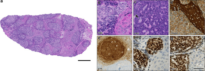
Fig. 2.
Morphological and immunohistochemical overview of the HPV-related multiphenotypic sinonasal carcinoma case 3 (HMSC-3). In a an overview is depicted with islands of larger basaloid cells in lobules with abundant mature squamous islands and keratinization. b Shows magnification of the larger, somewhat brighter basaloid cells with adjacent keratinization. In c a limited focus of a more conventional morphology with smaller, monomorphic basaloid cells and duct formation (arrowhead) is seen. d Shows pancytokeratin staining, visualizing the heterogeneous expression within the basaloid tumor cells and strong, diffuse positivity in the mature squamous islands. In e strong, diffuse expression of p16 can be seen in the basaloid tumor cells, whereas the squamous islands (lower right corner) lacks p16 expression. A similar pattern can be observed for SOX10 and LEF-1 (f, g). Scale bars 250 μm (a) and 50 μm (b–g)

