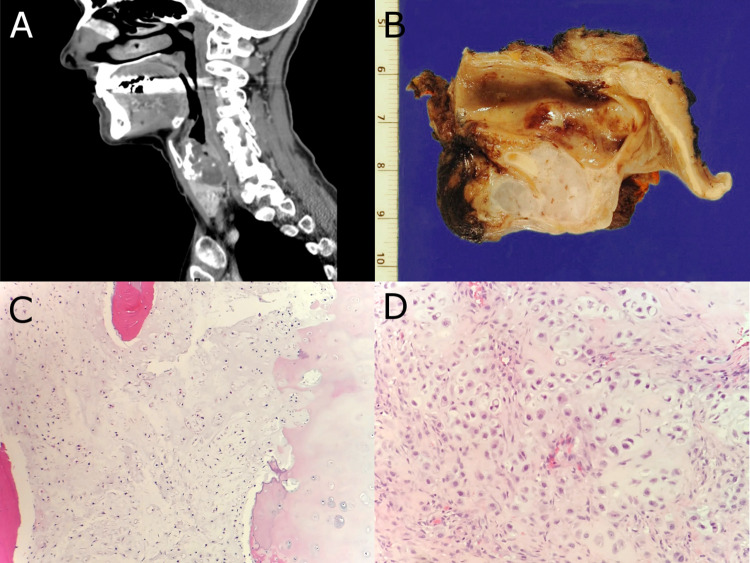
Fig. 1.
a, b Sagittal CT image and gross resection specimen from patient #2 demonstrating a destructive cartilaginous lesion which involves the cricoid cartilage. c H&E stained histologic section showing grade I conventional chondrosarcoma from patient #2. d H&E stained histologic section of grade II conventional chondrosarcoma from patient #3

