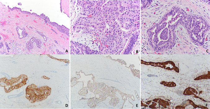
Fig. 2.
Secretory carcinoma of the salivary gland. Cyst wall with infiltrating glands showing focal cribriform and papillary architecture (a). Tumor cells are round, monomorphic, and have ample eosinophilic cytoplasm (b). Invasive gland surrounded by demosplastic stroma (c). Tumor cells are immunoreactive for S-100 (d), GATA3 (e), and mammaglobin (f)

