Fig. 2.

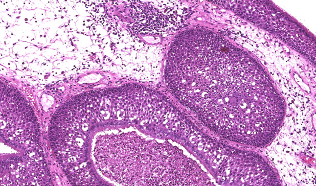
High power image shows non-keratinizing transitional epithelium covered by a layer of ciliated columnar epithelium. Infiltration by neutrophils is seen (hematoxylin-eosin 10x)

High power image shows non-keratinizing transitional epithelium covered by a layer of ciliated columnar epithelium. Infiltration by neutrophils is seen (hematoxylin-eosin 10x)