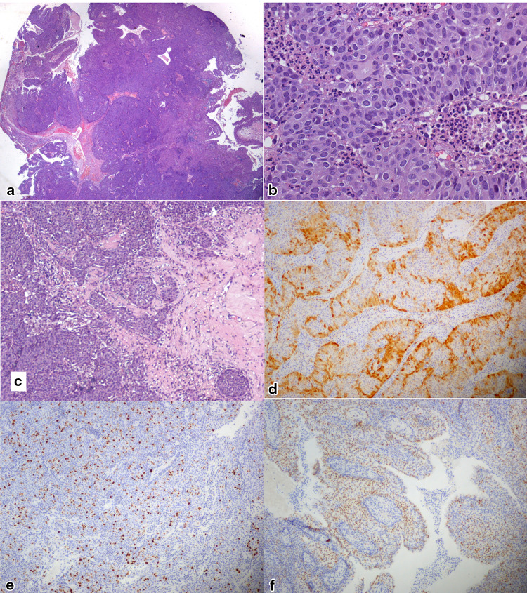
Fig. 1.
Photomicrographs documenting the morphologic and immunohistochemical features of LGPSC observed in Case 1: a low-power view (5 ×) of H&E-stained sections shows a complex papillary growth of epithelial neoplastic cells with broad, pushing nests; b high-power view (40 ×) of H&E-stained sections showing basaloid non-keratinizing cells with abundant cytoplasm, mitoses, and prominent granulocytic infiltrate; c neoplastic nests show deeply infiltrative behavior (10 ×); d P16 staining at the periphery of neoplastic cell cords and nests (10 ×); e MIB1-Ki67 was expressed in a variable proportion of neoplastic cells averaging 20% (10 ×); f P53 was expressed in 50% of neoplastic cells (10 ×)

