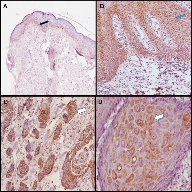
Fig. 4.
WNT5A expression by IHC in NOM, OED and OSCC. a Showing WNT5A in NOM (100X), weak focal positivity seen in membrane basal cell layers in few cases (black arrow). b Showing positive membraneous and cytoplasmic expression of WNT5A in dysplastic cell layers of OED (200X) (blue arrow). c Showing positive membraneous and cytoplasmic expression of WNT5A in dysplastic cells of tumor islands in OSCC (200X) (white arrow). d Positive membraneous and cytoplasmic expression of WNT5A in dysplastic cells within tumor islands in OSCC (400X)(white arrow)

