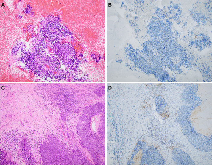
Fig. 3.
Case 6: a, b Cell block showing absent PD-L1 IHC staining of tumor cells and rare immunoreactive peritumoral immune cells. Incorporating findings in other microscopic fields, the overall CPS score was less than 1% (c, d). The corresponding hypopharyngeal tumor showed focal labeling of tumor cells as well as scattered clusters of immunoreactve peritumoral and intratumoral immune cells. The overall CPS score of this specimen was 2%

