Fig. 7.
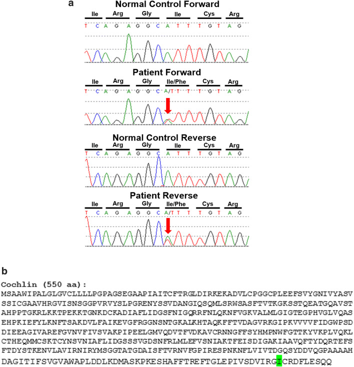
a Sanger sequencing traces, highlighting (red arrow) the c.1612A>T (p.Ile541Phe) variant in COCH. Sequencing was performed on both a control specimen and our patient in the forward direction (represented on the top) and in the reverse direction (on the bottom). The sequence traces from our patient show overlapping nucleic acid residues, adenine (A) and thymine (T), indicating heterozygosity for the variant in this position. b Cochlin amino acid sequence, showing the altered isoleucine (I) residue, highlighted in green

