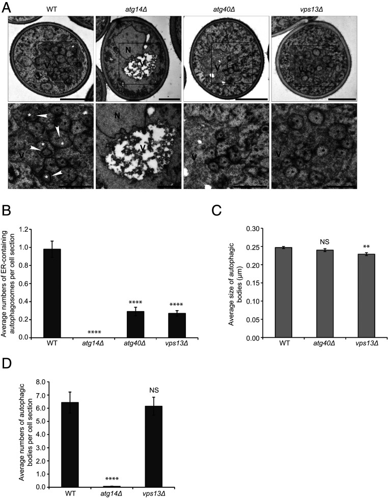
Fig. 6.
Packaging of the ER into autophagosomes is impaired in the vps13∆ mutant. (A, Top) Representative electron micrographs of yeast cells treated for 12 h with rapamycin. (A, Bottom) The boxed areas (Top) are enlarged. White asterisks mark an autophagic body containing an ER fragment; black asterisks mark an autophagic body lacking an ER fragment. Arrowheads point to an ER fragment inside an autophagic body. N, nucleus; V, vacuole. The darker tone of the vacuole lumen in WT, atg40Δ, and vps13Δ cells, relative to atg14Δ, is a consequence of continued autophagic flux in these strains. (B) Average number of ER-containing autophagosomes per cell section. (C) Average size of autophagic bodies. (D) Average number of autophagosomes per cell section. Error bars represent SEM; N = 100 cells. NS, nonsignificant (P ≥ 0.05), **P < 0.01, ****P < 0.0001, Student’s unpaired t test. (Scale bars, 1 μm [A, Top] and 500 nm [A, Bottom].)

