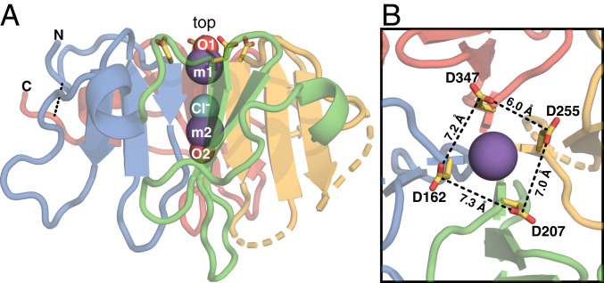
Fig. 1.
Structure of the Vn-HX propeller. Shown for molecule A of the asymmetric unit (Protein Data Bank ID code 6O5E). Backbone colors denote the repeat units corresponding to each propeller blade: HX1 (blue), HX2 (green), HX3 (yellow), and HX4 (red). Broken lines denote gaps in the protein chain due to missing electron density. The rim-Asp side chains (yellow/red) project from the top. (A) Side view. Spheres represent Na+ (m1 and m2; slate) and Cl− (aqua) ions occluded in the channel and water oxygen atoms (O1 and O2; red). The N- and C-termini are connected by a disulfide bond (dashed line). (B) Top view. The rim-Asp carboxylates form a ∼7 Å x 7 Å corral around the channel opening.

