Fig. 1.
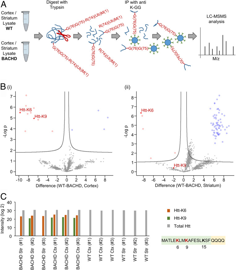
mHtt exon1 is highly and specifically ubiquitinated in HD. (A) Workflow for identifying ubiquitination targets and sites in cortical and striatal neurons from brains of BACHD and WT rats, following enrichment of ubiquitin-modified peptides by bead-conjugated anti–K-GG antibodies. R(74)(Ub)M(1) denotes the ubiquitin fragment (residues 1–74) released by trypsin. —G(76)G(75) denotes the two C-terminal Gly residues of ubiquitin remained bound in an isopeptide bond to an internal Lys residue in the target substrate. Details of the methods are described in the Ubiquitinome analysis of brain samples of the BACHD and WT rats section in Materials and Methods. (B) Volcano plots of differentially expressed ubiquitinated peptides from brain samples of WT and BACHD rats. The plots display significant results of a t test with permutation-based false discovery rate (FDR) calculation of fold change (difference) and significance (−Log p) using the Perseus software. (i) Visualization of ubiquitinated peptides from cortical brain samples. (ii) Visualization of ubiquitinated peptides from striatal brain samples. (C) Histogram showing intensities of Htt N-terminal peptides ubiquitinated on lysines 6 and 9 as well as total Htt measured in the same samples. Each sample is from the cortex or striatum of one WT or BACHD rat brain.

