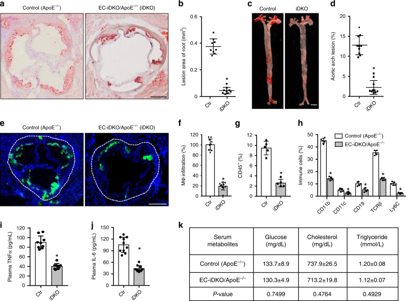
Fig. 5. Deletion of endothelial epsin 1 and 2 inhibits atherosclerosis.
a–d Oil Red O (ORO) staining of atherosclerotic lesions in aortic roots (a) and arches (c) of control (ApoE−/−) and EC-iDKO/ApoE−/− (iDKO) atherosclerotic mice fed a WD for 12–14 weeks. Statistical analysis of aortic root and arch lesions are shown in (b) and (d), respectively (n = 9 mice for aortic root, and n = 10 mice for aortic arch analysis, asterisk P < 0.001). Scale bars 500 μm (a) and 5 mm (c). e, f Macrophage (MΦ) infiltration in aortic roots by immunofluorescent staining with the Moma-2 antibody for control and iDKO mice fed a WD (e) and quantified (f) (n = 10 in each group, asterisk P < 0.001). Scale bar 500 μm. g Immune and inflammatory cells in aortic arches were measured using FACS analysis for CD45+ cells. Data are presented as percentage of the total cells analyzed (n = 5 in each group, asterisk P < 0.05). h Percentage of subtypes of immune cells in aortic arches measured by FACS for control and iDKO mice (n = 5 mice in each group, asterisk P < 0.001). i, j Serum TNFα and IL-6 levels in control and iDKO mice (n = 10 in each group, asterisk P < 0.001). k Metabolic parameters in serum (n = 10 in each group). Statistical analyses were performed using Student’s t-test and are presented as the mean ± SEM.

