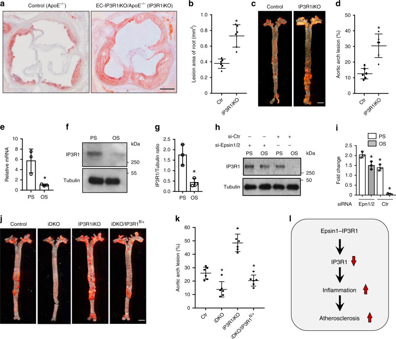
Fig. 7. Reduction of endothelial IP3R1 accelerates atherosclerosis.
a to d ORO staining of atherosclerotic lesions in aortic roots (a) and arches (c) of control (ApoE−/−) and EC-IP3R1iKO/ApoE−/− (IP3R1iKO) atherosclerotic mice fed a WD for 12–14 weeks. Statistical analysis of aortic roots and arches lesion are shown in (b, n = 6 aortic roots) and (d, n = 6 aortic arches), respectively (asterisk P < 0.001). Scale bars 500 μm (a) and 5 mm (c). e–g Human aortic endothelial cells (HAECs) subjected to 12 ± 4 dynes/cm2 pulsatile shear stress (PS) or 1 ± 4 dynes/cm2 oscillatory shear stress (OS) for 16 h. IP3R1 mRNA (e) and protein (f) levels were determined by qRT-PCR and western blot analysis, respectively. Quantitation of western blot results (g) (e–g, n = 3 independent repeats, asterisk P < 0.001). h,i HAECs subjected to PS or OS for 16 h in the presence of absence of epsin 1 and 2, followed by western blot (h) and statistical analyses (i) (epsin 1 vs. epsin 2, n = 3 independent repeats, asterisk P < 0.05; epsin 1 vs. Ctr, n = 3 independent repeats, asterisk P < 0.003; epsin 2 vs. Ctr., n = 3 independent repeats, asterisk P < 0.005;). j, k ORO staining of aortic arches of control, iDKO, IP3R1iKO, and iDKO/IP3R1fl/+ mice fed a WD for 26–28 weeks (j). Scale bar 500 µm. Quantification of ORO staining showing rescue of atherosclerosis progression in iDKO/IP3R1l/+ compared to iDKO mice (k) (n = 6 aortic arches in each group, asterisk P < 0.001). l Schematic diagram of endothelial epsin-mediated IP3R1 degradation and acceleration of inflammation and atherosclerosis. All data were assessed using Student’s t-test and are presented as the mean ± SEM.

