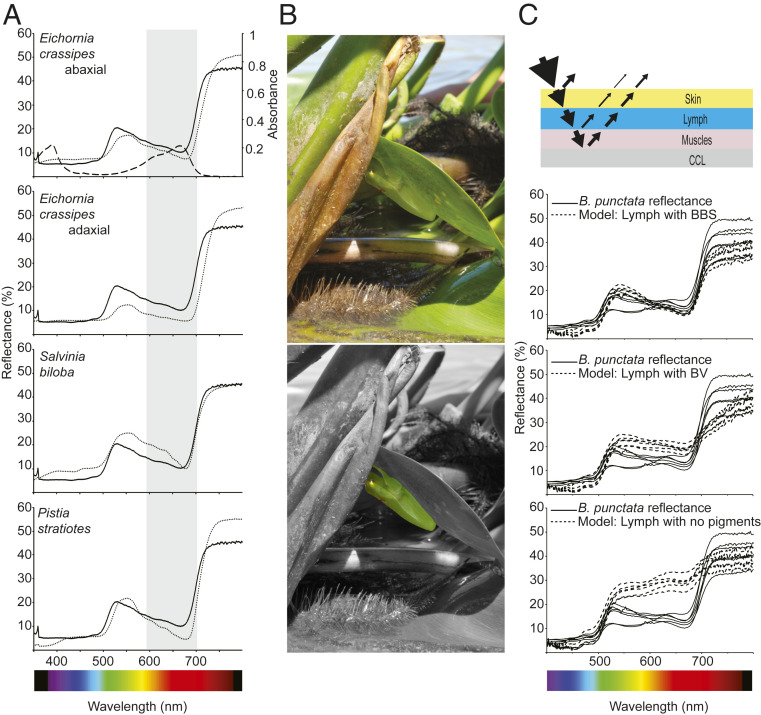
Fig. 4.
Influence of BBS in B. punctata coloration. (A) Reflectance spectra of frogs obtained from (2) (solid line) and different plants (dotted line) from their environment. BBS absorbance spectrum is shown in the Upper graph (dashed line), and both the shape and position of the Q band (shaded in gray) are consistent with the reduced reflectance of the animals in the red portion of the spectrum. Red-edge reflectance corresponds to a region of sharp change in reflectance at ∼695–700 nm, a phenomenon widely known in plants. The blue region is normally dominated by other pigments and can be modulated by fluorescence of hyloins (2). (B, Upper) Specimen of B. punctata in a normal resting position in the abaxial surface of a leaf of the water hyacinth Eichhornia crassipes during daytime. (B, Lower) Background is displayed in grayscale to highlight the frog position. (C) A simplified model of reflectance for the frogs to evaluate the influence of BBS in coloration. Light is reflected at the air skin interphase, transmitted through the translucent skins, and red and blue light are highly absorbed by lymphatic BBS. Under the lymphatic sacs, light is transmitted through the muscles and is reflected back in a broadband guanine-based reflector in the fascia (CCL) in the retroperitoneal cavity. Lymph can modulate the spectral properties of light emerging from the animals and it depends on its pigments’ composition. Physiological concentration of BBS exerts an important role in B. punctata coloration, and it outperforms the modeled effect in control experiments with isolated biliverdin (Middle graph) or with lymph devoid of BBS (Bottom graph). Solid lines: reflectance of six individuals. Dashed line: modeled reflectance for six independent skin samples.

