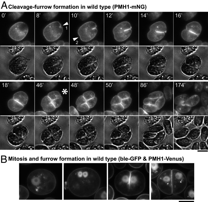
Fig. 1.
Live-cell observations of cleavage-furrow formation in Chlamydomonas. (A) Wild-type cells expressing the plasma membrane ATPase PMH1 tagged with mNG were synchronized using the 12L:12D/TAP agar method, mounted on TAP + 1.5% low-melting agarose, and imaged over several hours at ∼25 °C. Selected images are shown (times in minutes); the full series is presented in Movies S1 and S2. (Top) The mNG fluorescence (YFP channel); (Bottom) Differential-interference-contrast (DIC). Arrowheads, positions of the initial appearance of furrow ingression visible in this focal plane in the anterior (1) and posterior (2) poles of the cell. Asterisk, onset of second cleavage. (B) Wild-type cells coexpressing PMH1-Venus and the nuclear marker ble-GFP were imaged using a YFP filter set during growth on TAP medium at 26 °C. Cells at different stages in mitosis and cytokinesis are shown. (Scale bars, 5 µm.)

