Fig. 3.
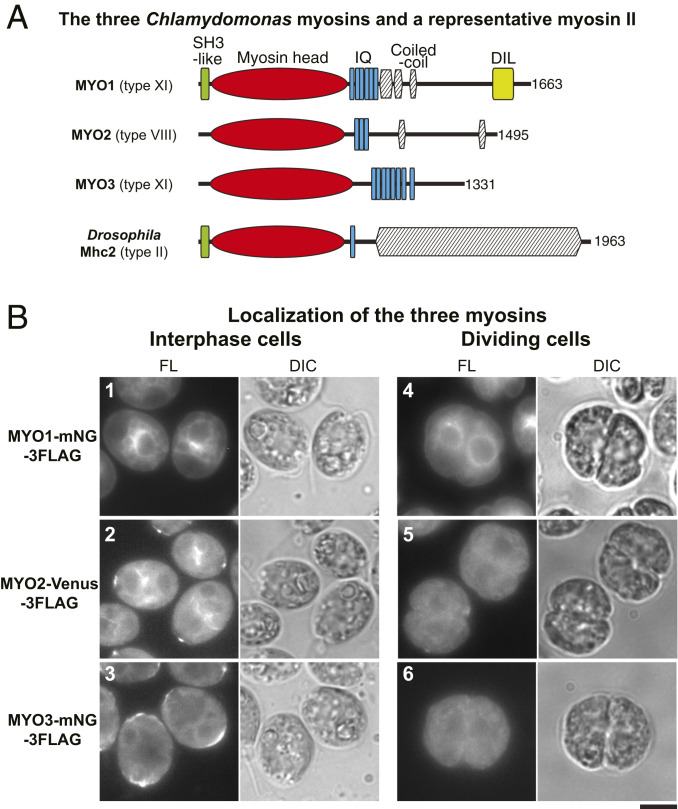
Lack of myosin localization to the region of the cleavage furrow. (A) Domain structures of the three Chlamydomonas myosins; a typical type-II myosin with a long coiled-coil tail (Drosophila Mhc2) is included for comparison. Domains were predicted using the HMMER (hmmer.org) and COILS (119) programs; total numbers of amino acids are indicated. (B) Localization of fluorescently tagged myosins in interphase and dividing cells; cells were grown on TAP medium at 26 °C. Fluorescence (FL) and DIC images are shown. (Scale bar, 5 µm.)

