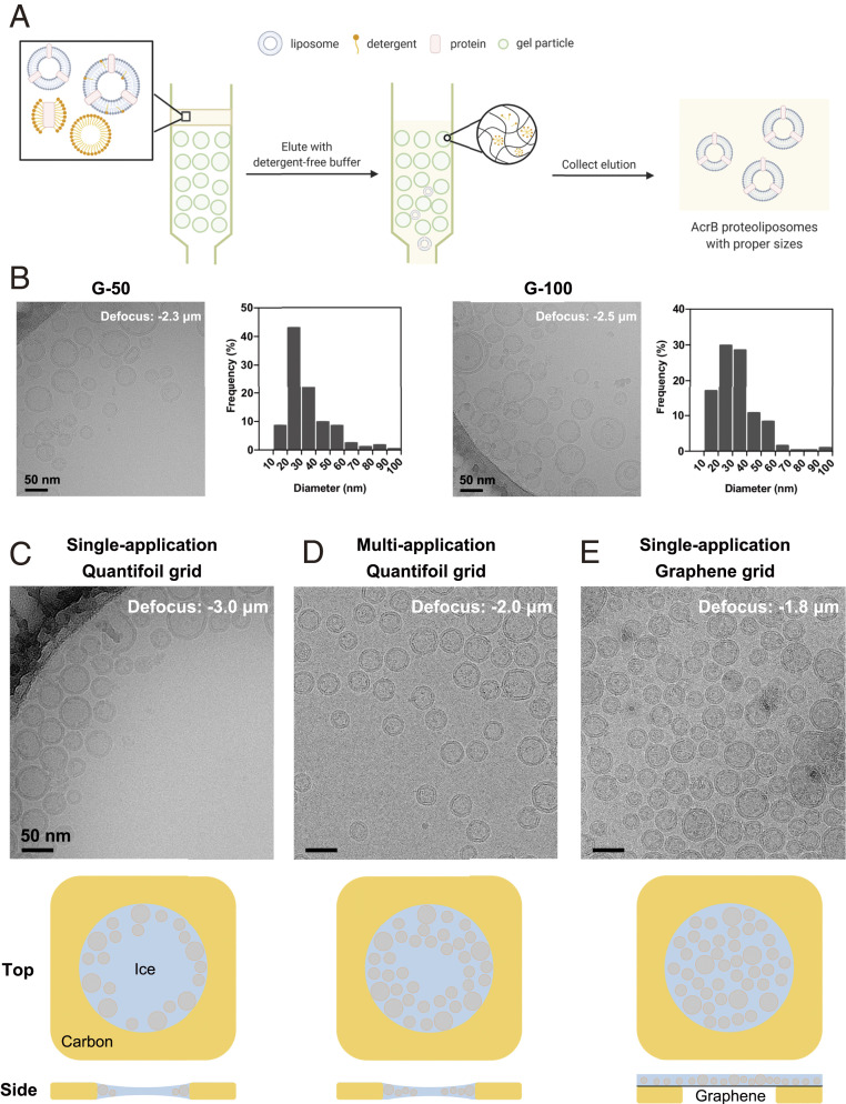
Fig. 1.
Optimization of proteoliposome cryo-sample preparation. (A) A schematic illustration of size selection of proteoliposomes using SEC. When the mixture of proteoliposomes and detergent micelles pass through different resins for SEC in the absence of supplemented detergents, liposomes are separated based on their diameters and detergents are removed. Please refer to SI Appendix, Fig. S1 (SI Appendix) for more trials of proteoliposome optimization. (B) Representative cryo-EM micrographs and size distribution of AcrB proteoliposomes prepared using manually packed Sephadex G-50 (Left) and G-100 columns (Right). Histograms are calculated from five holes of each sample. (C–E) Micrographs of AcrB proteoliposomes loaded on commercial Qauntifoil grids with (C) single- and (D) multiapplication approaches, and (E) on a graphene with single-application. The cartoon illustration for proteoliposome distribution in the holes by various sample preparation methods are presented below the corresponding panel.

