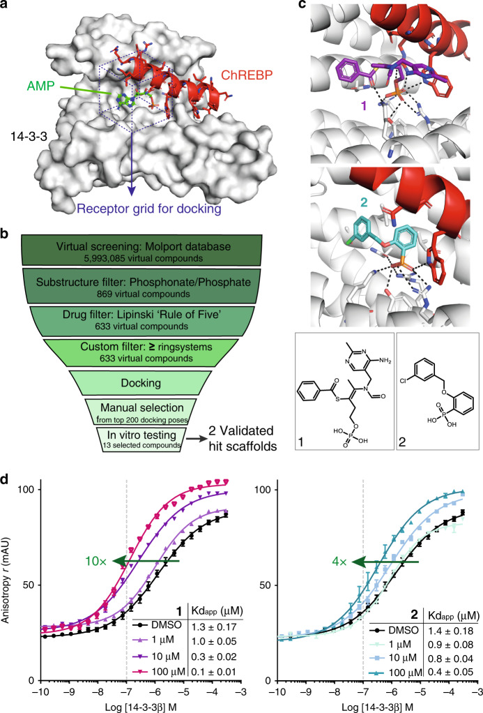
Fig. 1. A structure-based in silico screen for small-molecule stabilizers of the 14-3-3/ChREBP protein complex.
a The receptor grid (purple dotted box) for docking in the crystal structure of 14-3-3β (gray surface), ChREBP (red cartoon and sticks), and AMP (green sticks) (PDB entry 5F74)25. b Overview of the virtual screening procedure. c Docking poses and chemical structures of 1 and 2. d Titration curves of 14-3-3β on 100 nM fluorescently-labeled ChREBP peptide in the presence of increasing concentrations (1, 10, 100 μM) of 1 or 2. Data and error bars represent mean ± SEM, n = 3 replicates. Source data are provided as a Source data file.

