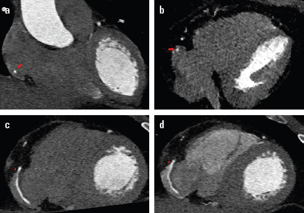
Figure 1.

. (a, b) Coronal image with cardiac synchronization and contrast only in left cavities which demonstrates the middle/distal third of the right coronary artery inside the right atrium (arrow). Plane images 4C oblique images following the axis of the artery. The intra-atrial path of the artery with contrast completely surrounding it. (c) With contrast in one phase. (d) With contrast in two phases to opacify the right cavities
