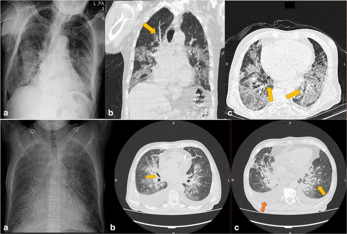
Fig. 1.
Upper row. Right a A 114-year-old male passed away from COVID-19 pneumonia. a Posterior anterior chest x-ray shows opacities in the lower lobes of both lungs. Center b Coronal CT image of the same patient, showing multilobar ground-glass opacities and septal thickening (yellow arrow). Left c Axial cut from the same patient shows multifocal ground-glass opacities and crazy-paving, characterized by a superimposed interstitial thickening on ground-glass opacities (yellow arrow). Down row. Right a A 60-year-old man passing away from COVID-19. a PA chest X-ray on admission, showing haziness in central regions. Center b An axial cut of the same patient. The yellow arrow marks an air bronchogram. Left c Axial cut showing reticulonodular opacities in both lungs, with the predominance of the left one (yellow arrow). This patient had a severe case of pleural effusion, most prominent on the right side (red arrow)

