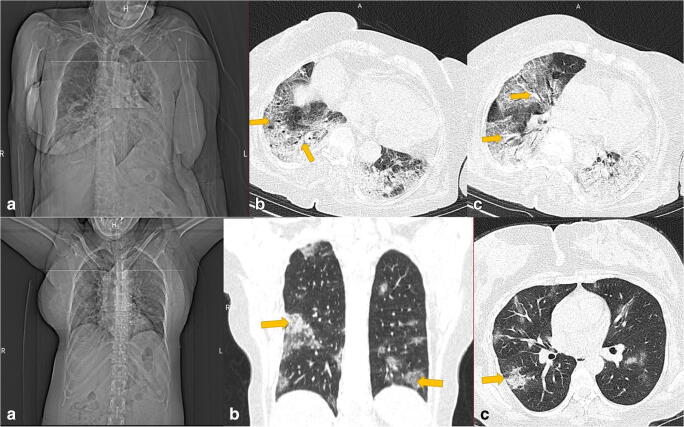
Fig. 2.
Upper row. An 83-year-old woman who expired from COVID-19. Right a PA chest x-ray of the patient, showing haziness and opacities in both lungs, predominantly the basal section of each lung. Center b Axial CT imaging showing atelectasis (yellow arrow) and diffuse involvement of both lungs. Left c Peribronchovascular involvement in the same patient. Down row. A 36-year-old female patient who survived COVID-19 and was hospitalized for 5 days. Right a PA chest x-ray of the patient. Center b Typical presentation of COVID-19, peripheral, multifocal ground-glass opacities (examples are shown by the yellow arrow). Left c Airspace consolidation is shown in the posterior segment of the right lung (yellow arrow)

