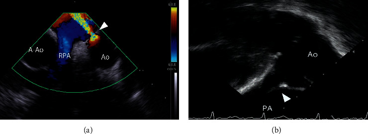
Figure 1.

Representative image of MPA and LPA views of PDA using ICE. (a) The MPA view shows the aortic short axis view. (b) The LPA view shows the PDA long axis. A‐Ao, ascending aorta; Ao, aorta; ICE, intracardiac echocardiography; LPA, left pulmonary artery; MPA, main pulmonary artery; PA, pulmonary artery; PDA, patent ductus arteriosus.
