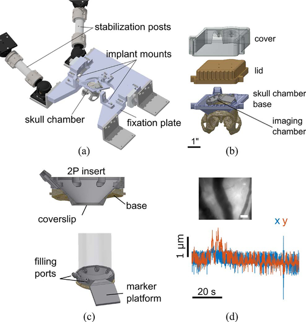
Figure 3:
Stabilization. (a) Head fixation and stabilization assembly. (b) Skull chamber assembly. (c) Imaging chamber assembly. The two photon insert seals to the chamber base using a gasket. (d) Motion artifact characterization. The top inset shows the reference image with which the motion artifact was quantified for the entire sequence. Scale bar: 100 μm. Blue and red traces show horizontal brain movement in the x and y dimensions, respectively.

