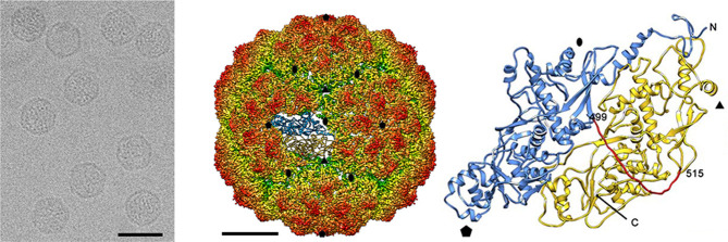
Fig. 1.
Structure of Penicillium chrysogenum virus. (Left) Cryo-EM image (bar, 50 nm), (Middle) Radially colour-coded three-dimensional cryo-electron microscopy reconstruction of the capsid viewed along a two-fold axis. The atomic structure of a monomer of the capsid protein is shown. Bar, 50 nm. (Right) Atomic model of capsid protein (top view) showing the N-terminal domain (1–498, blue), the linker segment (499–515, red) and the C-terminal domain (516–982, yellow). Symbols indicate icosahedral symmetry axes (adapted from [1]).

