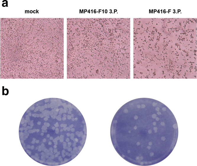
Fig. 1.

Virus isolation. (a) Photographs of mock-infected C6/36 cells and cells infected either with filtrated homogenate (MP416-F) or filtrated and 1 : 10 diluted homogenate (MP416-F10) 3 dpi. (b) Plaque morphology of the plaque-purified strain ASALV-PP in C6/36 cells 6 dpi.
