
Fig. 1.
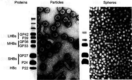
Negative-contrast electron micrographs of hepatitis B virus virions (left) and virus-associated particles (right; spheres are 17–22 nm in diameter), together with their SDS-PAGE protein profile. LHBs, MHBs and SHBs refer to large, middle and small hepatitis B virus surface proteins, respectively. HBc, hepatitis B virus core proteins. GP, glycoprotein; P, protein. (Courtesy of W. Gerlich).