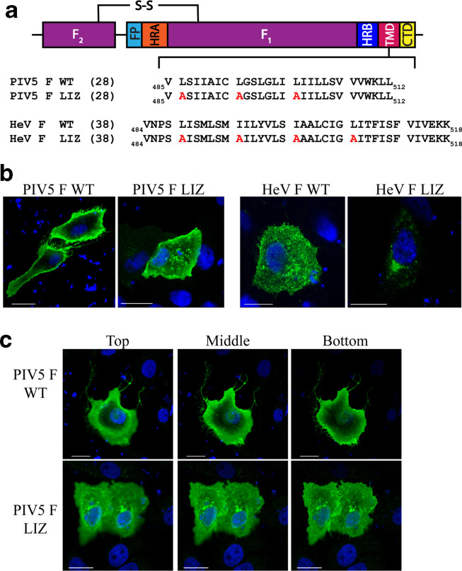
Fig. 1.
Mutations to the L/I zipper of HeV F and PIV5 F have variable effects. (a). Schematic of the paramyxovirus fusion protein highlighting the TM domain L/I zipper of HeV F and PIV5 F, and the mutant constructs. FP, fusion peptide; HRA, heptad repeat A; HRB, heptad repeat B; TMD, transmembrane domain; CT, cytoplasmic tail); S–S, disulfide bond. (b). Immunofluorescence to visualize localization of HeV and PIV5 F proteins. Vero cells were seeded in eight-well chamber plates and transfected with 0.75 µg PIV5 F WT or LIZ mutant (left), and HeV F WT or LIZ mutant (right). Localization of HeV F was analysed with anti-F 5G7 antibodies, and PIV5 F analysed with mAb F1a (green). Images were taken with a Nikon 1A confocal microscope. Images are representative. Scale bars represent 10 µm. (c). Z-stack images from (b) were collected in 0.3 µm sections, and images corresponding to top, bottom and middle slices are shown. Images are representative of two independent experiments carried out in triplicate. Scale bars represent 10 µm.

