
Fig. 1.
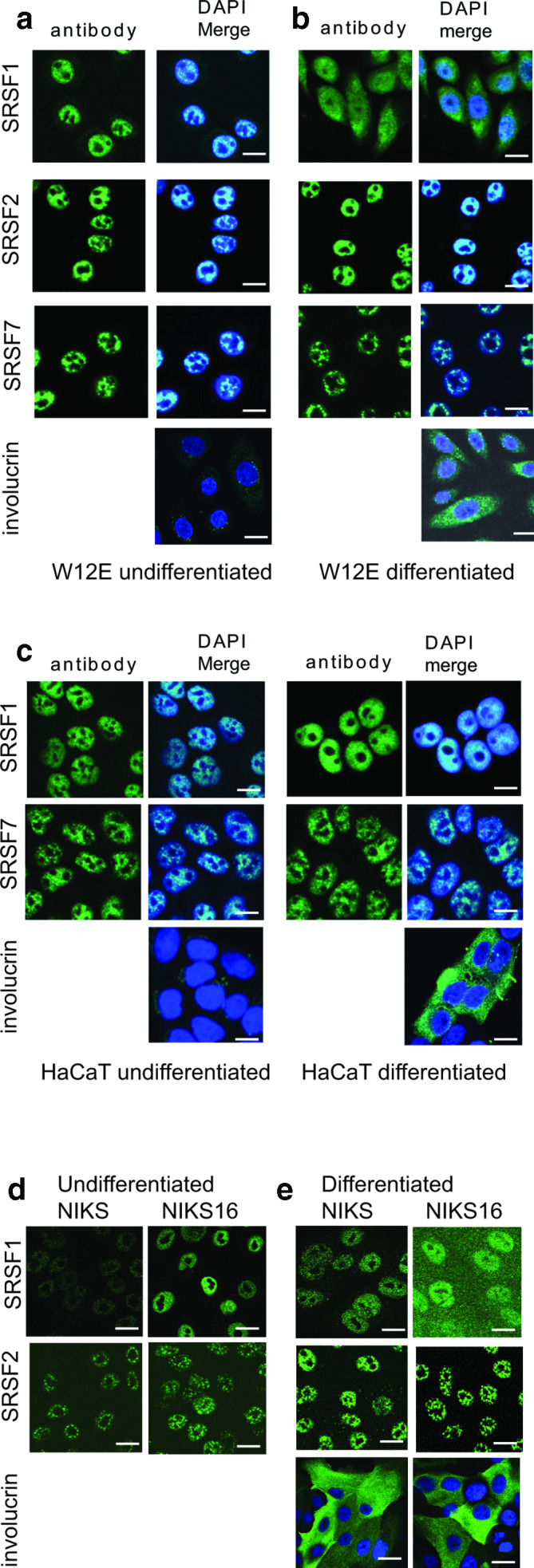
SRSF1 is present in the cytoplasm in differentiated HPV-infected keratinocytes. (a) Confocal microscopy analysis of the location of SR proteins SRSF1, SRSF2 and SFSR7 in undifferentiated W12E (HPV16-infected) keratinocytes. (b) Analysis of the location of SR proteins SRSF1, SRSF2 and SFSR7 in differentiated W12E (HPV16-infected) keratinocytes. (c) Analysis of the location of SRSF1 and SFSR7 in undifferentiated and differentiated HaCaT keratinocytes (HPV-negative). (d) Analysis of the location of SRSF1 and SRSF2 in undifferentiated NIKS (HPV-negative) and NIKS16 (HPV16-infected) keratinocytes. (e) Analysis of the location of SRSF1 and SRSF2 in differentiated NIKS (HPV-negative) and NIKS16 (HPV16-infected) keratinocytes. Cells were stained with involucrin to show differentiation. No involucrin staining was detected in undifferentiated cells. Nuclei were counterstained with DAPI (DAPI Merge). Bar, 20 µm.