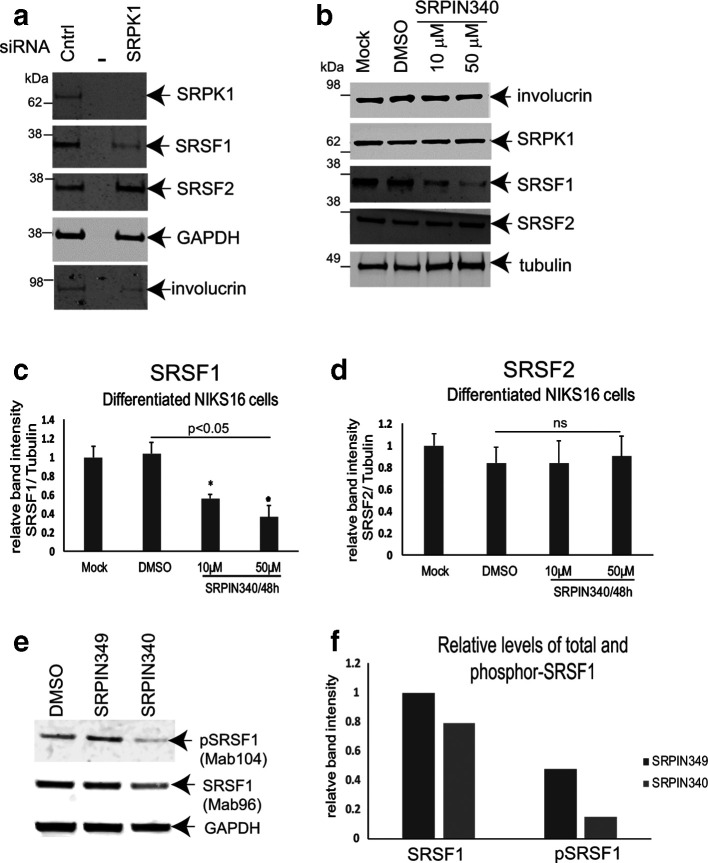
Fig. 5.
SRPK1 is required for SRSF1 phosphorylation in differentiated HPV16-positive keratinocytes. (a) Western blot analysis of total cellular SRSF1 levels (Mab96) in NIKS16 (HPV16-infected) cells treated with a control siRNA (Cntrl) or upon siRNA depletion of SRPK1 (SRPK1). The middle lane of the three is blank (–). GAPDH was used as a loading control. SRSF2 is shown as a control for an SR protein that is not significantly phosphorylated by SRPK1. Involucrin is shown as a control for keratinocyte differentiation. (b) Western blot analysis of total SRSF1 levels (Mab96) in NIKS16 cells untreated (Mock) or treated with the SRPK1 inhibitor SRPIN340 (10 µM, 50 µM) or with the vehicle, DMSO. SRPK1 levels are unaffected by drug treatment. SRSF2 is shown as a control for an SR protein that is not significantly phosphorylated by SRPK1. Involucrin is shown as a control for keratinocyte differentiation. Tubulin was used as a loading control. (c) Graph of relative levels of SRSF1 in untreated, DMSO-treated and SRPIN340-treated NIKS16 cells. (d) Graph of relative levels of SRSF2 in untreated, DMSO-treated and SRPIN340-treated NIKS16 cells. Western blot analysis of total (Mab96) and phosphor-SRSF1 (Mab104) levels in differentiated NIKS16 keratinocytes treated with drug vehicle, DMSO, or 10 µM SRPIN340 or SRPIN349, a similar compound that can bind SRPK1 but cannot inhibit its activity. (f) Quantification of the data in (e). The data are representative of two separate experiments.

