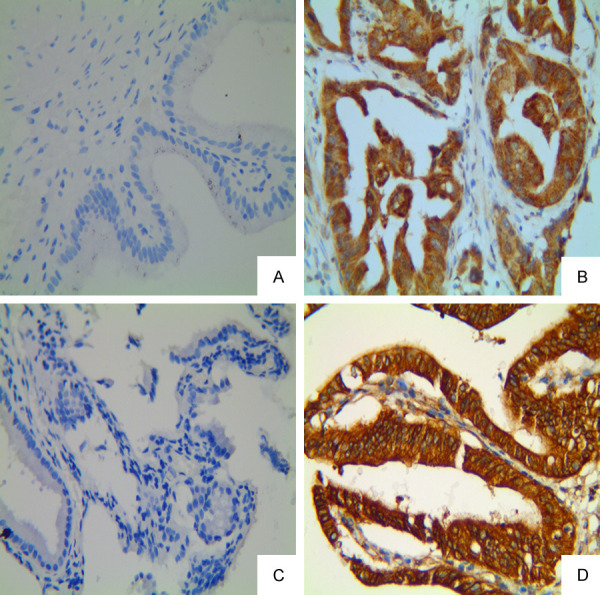
Figure 1.

Expression of FMNL2 and CTTN in GBAC and normal gallbladder tissue (Max Vision, original magnification: ×400). A, B: Negative staining of FMNL2 in the normal gallbladder tissue and positive staining in the cytoplasm of GBAC, respectively; C, D: Negative staining of CTTN in the normal gallbladder tissue specimen and positive staining in the cytoplasm of GBAC, respectively.
