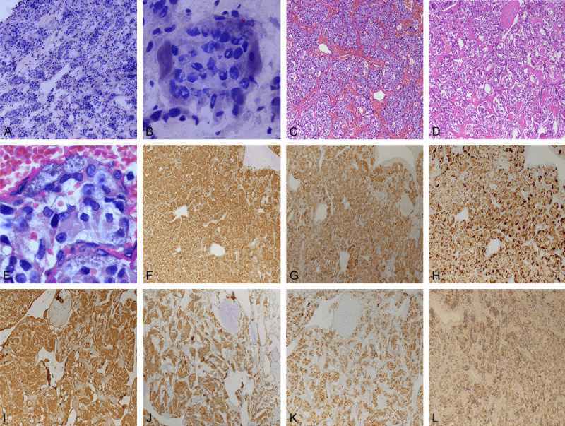
Figure 2.

(A) Low power image of frozen pathologic section of tumor (4×). (B) Clear sertoli cells could be seen around the main cell nests in frozen pathological section (40×). (C) Low power image of conventional pathologic section of vascular rich area in stroma of tumor (4×). (D) Low power image of conventional pathological section of sclerotic area in stroma of tumor (4×). (E) Sertoli cells could be seen around the main cell nests in conventional pathologic section (40×). (F) Main cells in (C) with positive expression of CgA by immunohistochemistry. (G) Main cells in (C) with positive expression of Syn by immunohistochemistry. (H) More or less sertoli cells strongly positive for S-100 protein around the main cell nests in (C). (I) Main cells in (D) with positive expression of CgA by immunohistochemistry. (J) Main cells in (D) with positive expression of Syn by immunohistochemistry. (K) Few or no sertoli cells strongly positive for S-100 protein around the main cell nests in (D). (L) Immunohistochemical positive expression of SDHB in tumor cells.
