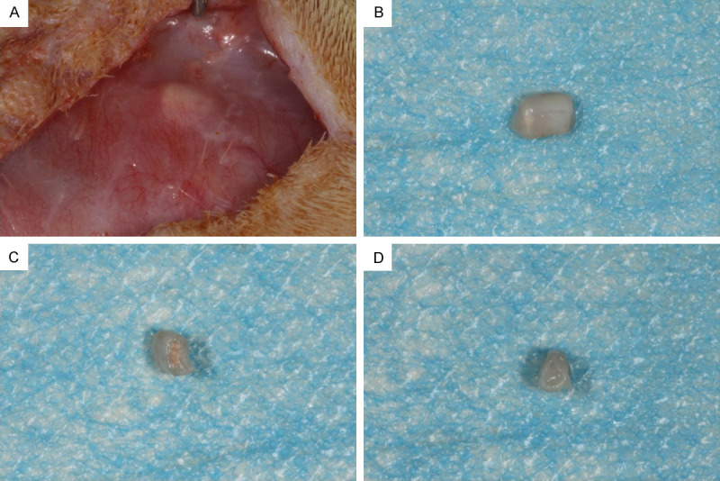
Figure 2.

General observations of the root fragments after the 3-month implantation. A. The blood supply radiated around the tooth root. B. When the specimen was removed, the surface of the specimen was surrounded by a transparent tissue membrane. C. In the rBMSC+iRoot BP group, new tissue filled the root segment, and some red blood vessels covered the side of the BP. D. In the control group, the pulp cavity was filled with pink or red tissues with a significant number of blood vessels on both sides of the root segment.
