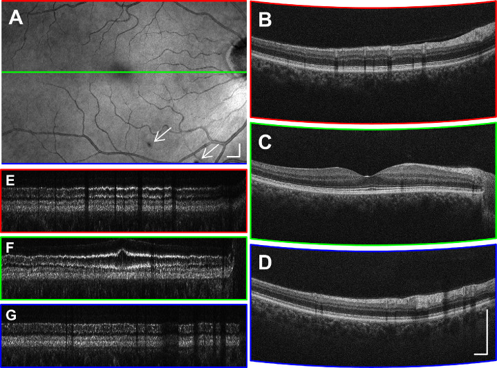
Figure 7.
Representative HD raster scan from a 58-year-old healthy participant. (A) The en face projection suggests that the B-scan spacing was adequate for detecting focal shadows (arrows). (B–D) A small amount of intra–B-scan ReALM was applied to flatten the retinal contour without producing excessive signal loss from fringe washout effects. (E–G) The BM-flattened 200-um slabs shown in linear display. Scale bars: 500 µm.

