
Figure 2. HFD-induced MDSC protect against metabolic dysfunction and reduce inflammation, but promote adiposity.
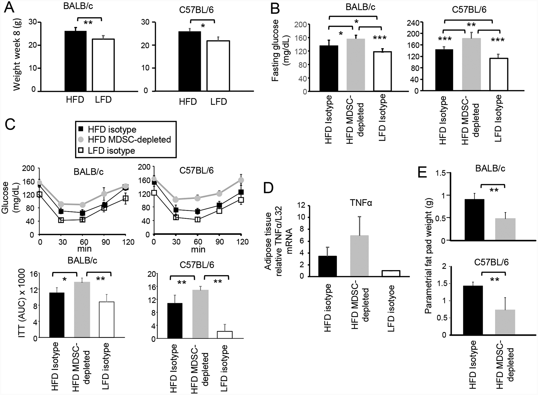
BALB/c and C57BL/6 female mice were maintained on a HFD or LFD diet for eight weeks. Some groups were concurrently depleted for MDSC or treated with isotype control antibodies. (A) HFD increases body weight. Mice on a HFD or LFD for 8 weeks were weighed. BALB/c: HFD n=20, LFD n=10; C57BL/6: HFD n=5, LFD n=5. (B) MDSC protect against HFD-induced elevated serum glucose. BALB/c and C57BL/6 mice on a HFD or LFD for 8 weeks were fasted for 6 hr and then tested for glucose levels in the blood. BALB/c: HFD and LFD n=10 mice/group; C57BL/6: HFD n= 5 mice/group, LFD n = 4 mice/group. (C) MDSC protect against insulin tolerance. Mice were injected with insulin, bled at 30 minute intervals, and the blood tested for glucose (insulin tolerance test). Area under the curve (AUC) values are pooled for 5 mice/group for each strain. (D) MDSC reduce inflammation in adipose tissue. RNA was isolated from the parametrial fat pads of BALB/c mice and assayed by qRT-PCR for expression of TNFα. Data are fold-change in HFD tissue relative to the same tissue from LFD mice. n=5 mice/group for HFD and LFD mice, and n=4 mice/group for HFD MDSC-depleted group. Data are from one of two independent experiments. (E) MDSC promote the accumulation of visceral adipose tissue. BALB/c and C57BL/6 HFD mice were MDSC-depleted or treated with control isotype mAb and their parametrial fat pads were dissected and weighed. n=4–5 mice/group.