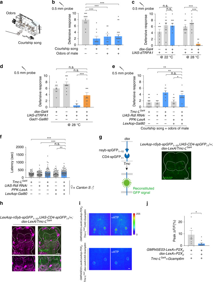
Fig. 2. Male cues suppress defensive response by activating dsx neurons.
a Schematic of simulative courtship assay. Tethered virgin flies were placed on top of a speaker playing back courtship song and 1 cm away from the chamber with males. Virgin flies were challenged with mechanical stimuli on their wing margin. b Wild-type (w1118) virgins placed either in the proximity of a group of male flies or a speaker playing fly courtship song had decreased defensive response. n = 13, 10, 8, and 19 for each group. ***p < 0.001, two-tailed unpaired t tests. c Activating dsx neuron with dTrpA1 in decapitated virgins caused decreased defensive response against mechanical stimuli. n = 9, 10, 11, 10, 10, and 10 (from left to right). ***p < 0.001, p > 0.05 (n.s.), two-tailed unpaired t tests. d Activating dsx neurons that do not release GABA in decapitated flies did not suppress defensive response. n = 9, 11, 9, and 10 for each group. ***p < 0.001, p > 0.05 (n.s.), two-tailed unpaired t tests. e Knocking-down GABAA receptor in all Tmc-L neurons or only CTNs with Rdl-RNAi did not suppress defensive response to mechanical stimuli in the virgin female flies exposed to courtship cues. n = 15, 8, 8, 8, and 10 for each group. **p < 0. 01, *p < 0. 05, p > 0.05 (n.s.), two-tailed unpaired t tests. f Knocking-down GABA receptor in all Tmc-L neurons or only CTNs with Rdl-RNAi in virgins prolonged time to copulation, and these virgins were paired with naïve Canton S male. n = 58, 55, 58, 57, 55 for each group. ***p < 0.001, **p < 0. 01, p > 0.05 (n.s.), two-tailed Mann–Whitney nonparametric test. g GRASP signal (green) between dsx neurons and CTNs in female VNC highlighted with white dashed line. White line outlined the border of the mesothoracic ganglion of the VNC. Scale bar represented 50 μm. h GRASP signal (green) between dsx neurons and CTNs in female VNC was positive to GABA immune-staining (magenta). GRASP signal was highlighted with white dashed line. GRASP signal region was zoomed in below. Scale bar represented 50 μm (upper) and 25 μm (lower). i Activation of wing mechanosensory neurons via P2X2 triggered a robust Ca2+ influx in CTNs’ cell bodies (upper). The influx was inhibited when wing margin mechanosensory neurons and dsx neurons were activated simultaneously (lower). The cell bodies of CTNs were cycled with white dashed lines. Scale bar represented 50 μm. j Peak fluorescence changes (ΔF/F) of CTNs’ cell bodies when activating wing margin mechanosensory neurons (left bar, n = 6), or activating wing margin mechanosensory neurons and dsx neurons simultaneously (right bar, n = 10). *p < 0. 05, two-tailed Mann–Whitney nonparametric test. Experiments in d–f, i, j) were done with virgins. Error bars indicate mean ± SEM, n.s., not significant. Source data are provided as a Source Data file.

