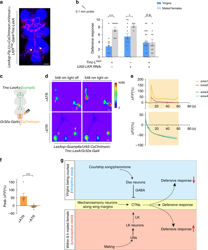
Fig. 6. CTNs are activated by LK to induce increased defensive response in mated females.
a Intersection between Tmc neurons and LKR neurons (red). White arrow indicated the cell bodies of CTNs. Blue: nc82. Scale bar represented 50 μm. b Knocking-down LKR in Tmc-L neurons blocked the mating-induced increase of defensive response. n = 11, 10, 13, 6, 20, and 11 (from left to right). ***p < 0.001, *p < 0.05, p > 0.05 (n.s.), n.s., two-tailed unpaired t tests. c Schematic for assays in (d). d Representative imaging of Ca2+ responses in CTNs’ cell bodies in the VNC before (left) and after (right) activating UNs with CsChrimson. Upper panels: flies fed with all-trans retinal (ATR). Lower panels: without ATR. The cell bodies of CTNs were cycled with white dashed lines. Numbers indicated different cell bodies. Scale bar represented 50 μm. e Representative traces of ΔF/F of CTNs’ cell bodies showed in (d) after UNs activation. f Ca2+ signal of flies supplied with ATR (left bar, n = 6) was elevated by UNs stimulation compared with flies without ATR (right, n = 12). ***p < 0.001, two-tailed Mann–Whitney nonparametric test. g Working model for courtship and mating mediated behavioral switch of defensive response in female flies. Error bars indicate mean ± SEM, n.s., not significant. Source data are provided as a Source Data file.

