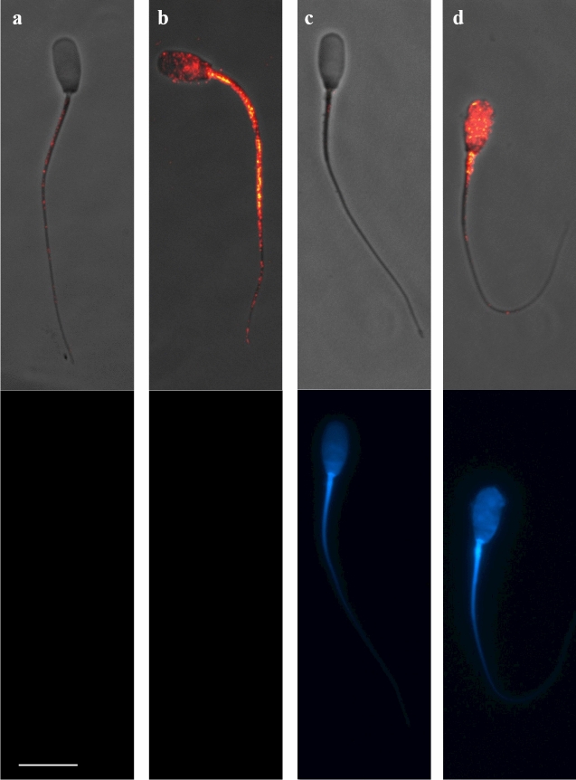
Figure 4.
Immunocytochemistry of GM-CSF in the pig mature spermatozoa. Top images (a and c) show spermatozoa with low/absent GM-CSF expression and bottom images show spermatozoa viable and non-viable (DAPI stain). The sperm samples were incubated with anti-GM-CSF antibody (orb6090, Biorbyt) to identify GM-CSF expression (composite in red in top images) and incubated with DAPI staining to identify viable from non-viable (DAPI positive in blue in bottom images) spermatozoa. Sperm in (a) and (c), were included in cluster 1 (fluorescence intensity 4.45 ± 0.17 RU; ranging from 0.0 to 15.33 RU) while spermatozoa displaying high GM-CSF expression were included in cluster 2 (fluorescence intensity 29.81 ± 1.82 RU; ranging from 15.67 to 84.0 RU, b and d). Scale bar: 10 μm. Cropped image of immunocytochemistry capture (see Supplemental Information, Fig S5 for full image).

