Fig. 2.
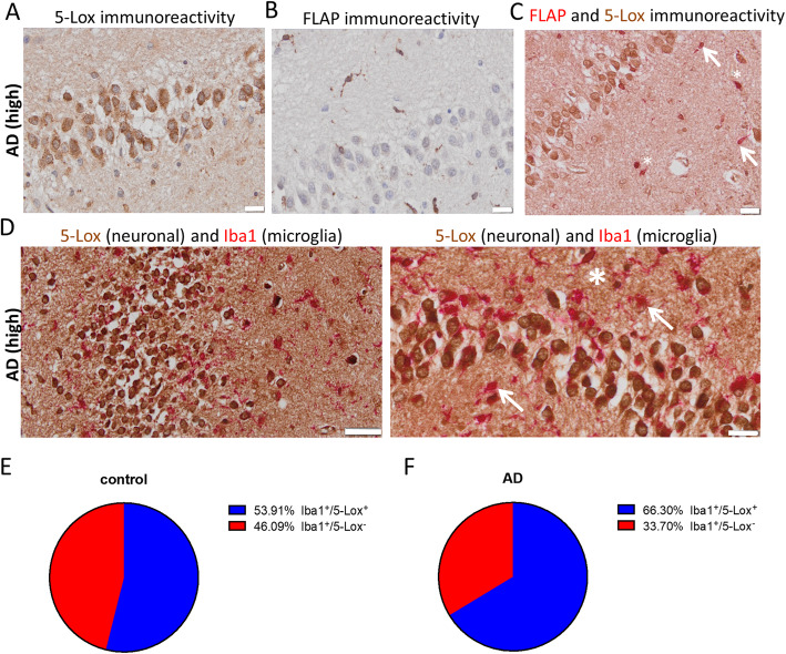
Immunohistochemical analysis for 5-Lox and FLAP expression in human AD brain. a 5-Lox immunoreactivity (using monoclonal 5-Lox antibody from BD Biosciences #610694) in human post-mortem AD (high, Braak 6) hippocampus specimen. 5-Lox staining was prominent in cells of the granular cell layer in the dentate gyrus. b FLAP was expressed in cells with glial cell morphology but not in neurons. c Double staining of 5-Lox with FLAP revealed only minor co-expression of 5-Lox in FLAP positive cells (arrows). Asterisks indicate double positive cells. d Double staining of 5-Lox with microglia marker Iba1 showed that non-neuronal 5-Lox expression is associated to some microglia cells (asterisk). Arrows indicate microglia without 5-Lox expression. e Percentage of Iba1+/5-Lox+ and Iba1+/5-Lox− cells in Iba1+ cells from control and AD (f) hippocampus sections. Hematoxylin (a, b) was used as nucleus stain. Scale: 20 μm (a-c, d right image), 50 μm (d, left image)

